咨询电话:
400-6018-657


 AddTime:2022-01-07
Clinical application range:
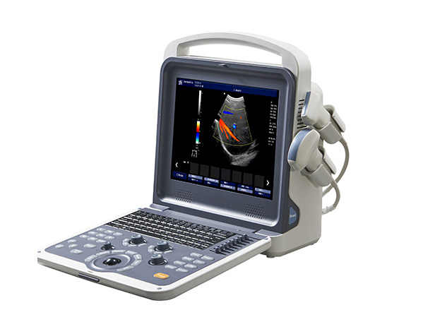
Used for clinical ultrasound diagnosis. Mainly for diagnosis and analysis of human abdominal organs (liver, gallbladder, pancreas, spleen, kidney), obstetrics and gynecology, thyroid, small organs, urology, superficial, orthopedic surgery. , Carotid artery, intestine, breast and other parts of the bloodstream for ultrasound diagnosis and inspection.

Feature:
Clinical application range:
Used for clinical ultrasound diagnosis. Mainly for diagnosis and analysis of human abdominal organs (liver, gallbladder, pancreas, spleen, kidney), obstetrics and gynecology, thyroid, small organs, urology, superficial, orthopedic surgery. , Carotid artery, intestine, breast and other parts of the bloodstream for ultrasound diagnosis and inspection.
Performance characteristics:
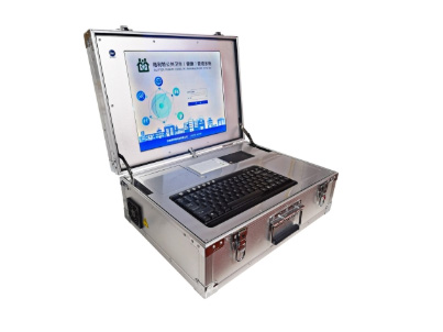
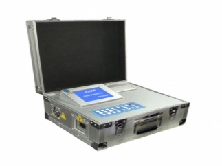
1. Portable design: portable clamshell structure, the net weight of the whole machine is less than 6 kg; twelve technical parameters can be adjusted independently with a single knob;
2. Test items: Diagnosis and analysis of human abdominal organs (liver, gallbladder, pancreas, spleen, kidney), obstetrics and gynecology, thyroid, small organs, urology, superficial, orthopedic surgery, liver, gallbladder, pancreas Ultrasound diagnosis and examination of blood flow in the spleen, kidney, carotid artery, intestine, breast, etc .;
3. Display control: reverse display, zero shift, B-refresh, D expansion, Doppler angle correction, B / D expansion, partial discharge and shift;
4. Display mode: speed dispersed display, speed display, energy display;
5. Ultrasonic power output adjustment: B / M, PWD, CF output power can be adjusted ≥ 8 levels; spectrum false color, spectrum reverse function;
6. Can be interconnected with the information terminal equipment, the test results can be passed in at any time.
Technical specifications:
1. Basic description of system general functions:
1.1 Display: high-resolution wide-screen LCD monitor, image resolution ≤0.5mm, can display hairline;
1.2 Full digital color ultrasound diagnostic system host, high-resolution blood flow imaging technology;
1.3 Two-dimensional gray-scale imaging unit, speckle noise suppression technology, high-fidelity imaging technology, all-digital beamformer;
1.4 Color Doppler blood flow imaging unit, energy Doppler blood flow imaging unit, directional energy Doppler blood flow imaging unit, PW Doppler blood flow imaging unit, M-type imaging unit;
1.5 Tissue optimized imaging technology, iclear programmed scanning technology, adaptive color artifact removal technology, automatic spectrum tracking measurement, 3K-10K extended pulse imaging technology, and multiple signal parallel processing technology;
1.6 The dynamic range of the host is ≥150dB, visually adjustable from 0 to 150dB;
1.7 Portable clamshell structure, the net weight of the whole machine is less than 6 kg; the twelve technical parameters can be adjusted independently with a single knob.
2. Probe specifications: optional convex array, linear array, endoscopic probe and other probes, and can be used in conjunction with the metal puncture frame;
3. Main parameters of two-dimensional image:
3.1 Focusing: the whole screen and the whole area of the transmitting focus area, and the digital dynamic receiving focus in the whole process;
3.2 Total gain adjustment ≥100dB, B / M / D / CFM can independently adjust the gain, STC segment ≥8;
3.3 Maximum image depth ≥280mm, system dynamic range ≥130dB, gray-scale imaging display ≥128 level;
3.4 PSHI TM broadband frequency shift, optimized tissue harmonic imaging;
3.5 Magnification function: panoramic magnification and partial magnification, the magnification level ≥ 10 levels;
3.6 Preset mode: According to different inspection organs, the inspection of optimized images can be preset,
3.7 Preset quantity: ≥8 kinds, users can customize the conditions.
3.8 One-key full-screen function of ultrasound image.
4. Spectrum Doppler;
4.1 Mode: Pulse wave Doppler (PWD);
4.2 Sampling width and position range: width 0.5mm to 48mm graded adjustment;
4.3 Angle correction: -80- + 80 degrees, the incident angle is between 45-60 degrees;
4.4 Display control: reverse display, zero shift, B-refresh, D expansion, Doppler angle correction, B / D expansion, partial discharge and shift;
4.5 Display mode: speed dispersion display, speed display, energy display;
4.6 Ultrasonic power output adjustment: B / M, PWD, CF output power can be adjusted ≥ 8 levels; spectrum false color, spectrum reverse function.
5. Color doppler
5.1 Imaging methods: color Doppler velocity map, color Doppler energy map, directional energy map;
5.2 Automatic color technology: single-step operation, one-click optimization of color Doppler blood flow;
5.3 Scanning rate: full field of view, frame rate ≥50 frames / second at 18cm depth;
5.4 Display format: double-frame real-time display B + B / C mode, double-frame display, B / C mode,
B / C / Doppler triple synchronization or dynamic refresh mode;
5.5 The angle change range of linear array probe scanning imaging is ≥20 degrees;
5.6 The blood flow box can be inverted.
6. System measurement and analysis:
6.1 General measurement: distance, area, perimeter, volume, angle, time, slope, flow rate, flow rate ratio, etc .;
6.2 Doppler blood flow measurement and analysis, peripheral blood vessel measurement and analysis.
7. Image storage, management, recording and movie playback reproduction unit:
7.1 The system supports B, M, CF, PW images, and supports B / M, B / PW movie playback;
7.2 Number of movie playback frames ≥1000 frames;
7.3 Automatic entry of user information;
7.4 Dynamic images, static images, direct storage, can directly view images on ordinary PC, image instant printing technology; video and report storage copy function;
7.5 Built-in battery can last 2 hours, AC and DC automatic conversion.
8. Input / output signal:
8.1 Input: COM, USB, DICOM; Output: VGA, DVI, USB, COM, DICOM;
8.2 Connectivity: With DICOM3.0 interface software package for medical digital images and communication;
9. Data transmission: can be interconnected with the information terminal equipment, the test results can be passed in at any time.
ADVANTAGE OF THE SERVICE
The powerful processing capability of the domain optical platform, high frame rate shear wave elasticity can achieve higher two-dimensional elastic shear wave imaging speed and display frame rate, improve the temporal resolution of the elastic images, and have more confidence in clinical diagnosis.


The powerful processing capability of the domain optical platform, high frame rate shear wave elasticity can achieve higher two-dimensional elastic shear wave imaging speed and display frame rate, improve the temporal resolution of the elastic images, and have more confidence in clinical diagnosis.


The powerful processing capability of the domain optical platform, high frame rate shear wave elasticity can achieve higher two-dimensional elastic shear wave imaging speed and display frame rate, improve the temporal resolution of the elastic images, and have more confidence in clinical diagnosis.

Wechat public account